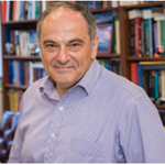

 Prof. Diomedes Logothetis
 My graduate training introduced me to the complex but fascinating world of molecular mechanisms of regulation of protein function. Two years after the patch-clamp technique was published, I joined David Clapham’s lab in 1983 and undertook as a PhD thesis project the mechanism by which the activity of K+ channels was regulated by G proteins. Perfusion of inside-out patches with purified G proteins revealed that the G subunits of G proteins were sufficient to stimulate channel activity in a manner independent of the endogenous G proteins1. In my postdoctoral work I trained with the late Peter Hess and studied N-type Ca2+ channels and their regulation by G proteins2. In Bernardo NadalGinard’s lab I learned molecular biology and studied voltage-sensing mechanisms of K+ channels3. As a junior faculty in my own lab at Harvard Medical School, I also mentored an MD/PhD student in monitoring non-invasively the electrical activity of dissociated neurons from the suprachiasmatic nucleus that showed circadian activity patterns, an area I had studied in my Masters studies at Northeastern University and as a technician at Harvard Medical School before I started my PhD studies4.
I started my own lab in Manhattan at the Mount Sinai School of Medicine and returned to the study of G protein regulation of K+ channel activity, using structure and function relationships. Important contributions were made in the next decade5-8. Yet, an important paper 5 years into my career at Mount Sinai took us in a seemingly different direction, namely the dependence of G protein activation of K+ channels on phosphoinositides and PIP2 in particular9. PIP2 turned out to be a key regulator of the activity of most ion channels and my lab in 1998 found itself in an opportune position to aggressively pursue this area10-17. The coupling of the G protein regulation to the PIP2 regulation and the dependence of most channels on this minor phospholipid residing in the inner leaflet of the plasma membrane bilayer intrigued me.
In 2008, I accepted a new position at the Virginia Commonwealth University School of Medicine. The opportunity to build a strong Physiology and Biophysics department appealed to me and I have had great fun doing so, despite the grim funding times of science in the U.S.. My own laboratory program was to continue on the PIP2 and G protein regulation of ion channels. Yet, another important paper came in 2011 to stimulate my interest yet in a third direction. Using PIP2 and G protein-dependent ion channels expressed in Xenopus oocytes we realized we had a sensitive and reliable reporter of GPCR function. We set out to study a heteromeric GPCR involved in schizophrenia and the results were spectacular. The oocyte system and our assay was the best way to figure out empirically the ratio of the two receptors that yielded the most efficient cross signaling. With a clear assay, we managed to decipher the mechanism by which antipsychotic drugs worked through this heteromeric complex and tested the predictions of our model in native tissues and behavioral assays18. Thus heteromeric receptor function has become a third area of inquiry in my lab and one of great application to many physiological systems and pathophysiological conditions. The three current areas of inquiry in my lab are connected in a fundamental manner. I believe that these membrane-delimited molecules (channels, GPCRs, heterotrimeric G proteins, and PIP2) are working in concert as part of a macromolecular assembly. I am convinced that the only way to gain molecular understanding of their coordinated activities is to study their function in terms of their 3-dimensional structures.
I look forward to making significant contributions to this third research area that is nicely connected to my other lines of research, particularly to the structure-function understanding of the G protein signalosome.
1. Logothetis et al., 1987, see ref. #1 above, Nature 325: 321-6, 987 citations
2. Plummer, Logothetis and Hess, 1989, Neuron 2: 1453-63, 636 citations
3. Logothetis et al., 1992, Neuron 8: 531-40, 136 citations
4. Walsh et al., 1995, Neuron 14: 697-706, 1069 citations
5. Chen et al., 1995 – ref. #2, Science 268: 1166-9, 248 citations
6. Jin et al., 2002, Molecular Cell, 10: 469-81, 114 citations
7. He et al., 1999, J. Biol. Chem.274: 12517-24, 103 citations
8. He et al., 2002, J. Biol. Chem. 277: 6088-96, 95 citations
9. Sui et al., 1998 – ref. # 6, PNAS 95: 1307-12, 194 citations
10. Rohacs et al., 2005 – ref. #3, Nature neuroscience 8: 626-34, 380 citations
11. Zhang et al., 2003 – ref. #10, Neuron 37: 963-75, 325 citations
12. Lopes et al., 2002 – ref. #9, Neuron 34: 933-44, 290 citations
13. Zhang et al., 1999 – ref. #7, Nature Cell Biology 1: 183-8, 268 citations
14. Kobrinsky et al., 2000 – ref. #8, Nature Cell Biology 2: 507-14, 212 citations
15. Du et al., 2004 – ref. #12, Journal of Biological Chemistry 279: 37271-81. 147 citations
16. Rohacs et al., 1999, Journal of Biological Chemistry 274: 36065-72, 146 citations
17. Rohacs et al., 2002 – ref. #11, PNAS 100: 745-50, 136 citations
18. Fribourg et al., 2011 –ref. #4, Cell 147: 1011-23, 83 citations
G protein-sensitive inwardly rectifying K+ (GIRK) channels are activated by the subunits of G proteins via Gi/o-protein coupled receptors (Gi/oPCRs). GIRK channels utilize phosphatidylinositol 4,5-bisphosphate (PIP2) to maintain their activity. Hydrolysis of PIP2, induced by stimulation of Gq-coupled receptors (GqPCRs), reduces channel activity. We have utilized GIRK channels as reporters of GPCR function in the heterologous Xenopus laevis oocyte expression cell system by expressing receptor and channel cRNAs. Our studies of two heteromeric GPCR complexes, the 5-HT2A/mGlu2 and the 5-HT2A/D2, involved in schizophrenia have yielded unexpected results and revealed the unifying mechanisms of action of antipsychotic drugs.